
21-Day Fatty Liver Diet Plan: A Practical Guide to Better Liver Health

21-Day Fatty Liver Diet Plan: A Practical Guide to Better Liver Health If you have been diagnosed with fatty liver, one of the first questions you may have is, “What should I eat to reduce liver fat?” Searching for a 21 day fatty liver diet plan can be a useful starting point, but it is important to understand that there is no single three-week diet that can permanently “cure” fatty liver. The good news is that consistent dietary and lifestyle changes can help reduce excess liver fat and improve metabolic health. For people who are overweight or have obesity, gradual weight loss can reduce liver fat and may also improve inflammation and fibrosis. This 21-day plan is designed around practical principles that can be continued beyond three weeks. It emphasizes vegetables, whole grains, fiber-rich foods, lean protein and healthier fats while reducing added sugar, highly processed foods and excessive saturated fat. Medical note: This is a general educational diet framework, not an individualized prescription. People with diabetes, kidney disease, cirrhosis, pregnancy, significant weight loss, or other medical conditions may require a different nutritional plan. Consult a gastroenterologist, hepatologist or qualified dietitian before making major dietary changes. What Is Fatty Liver? Fatty liver occurs when excess fat accumulates in the liver. The condition is commonly associated with metabolic factors such as overweight or obesity, insulin resistance, type 2 diabetes and abnormal blood lipids. The terminology has also evolved. What was traditionally called non-alcoholic fatty liver disease (NAFLD) is now commonly referred to as metabolic dysfunction-associated steatotic liver disease (MASLD). Not everyone with fatty liver develops serious liver damage. However, in some people, ongoing inflammation can lead to fibrosis, cirrhosis and other complications. This is why identifying fatty liver early and addressing its underlying risk factors is important. Can a 21-Day Diet Plan Reverse Fatty Liver? A 21-day plan should be viewed as a starting point rather than a guaranteed cure. There is no scientifically established rule that fatty liver disappears after exactly 21 days. Liver health generally improves through sustained changes in diet, physical activity, weight management and control of conditions such as diabetes and high cholesterol. For people who need to lose weight, even modest weight loss can reduce liver fat. Maintaining a healthy weight over time can support liver health by helping reduce inflammation and the risk of progressive scarring. AASLD educational guidance notes that a 5–10% reduction in body weight is associated with meaningful improvements in several aspects of MASLD. Therefore, the purpose of this 21 day fatty liver diet plan is to help establish habits that can continue for months and years. Basic Principles of a Fatty Liver Diet Before starting the three-week plan, focus on these simple principles: Eat More Green leafy vegetables Cruciferous vegetables Fresh whole fruits Oats and other whole grains Brown rice and other minimally processed grains Dal and beans Fish Eggs Skinless chicken Low-fat or unsweetened dairy, when appropriate Nuts and seeds in controlled portions Healthy unsaturated fats Eat Less Sugary drinks Sweets and desserts Bakery products Deep-fried foods Processed snacks Excessively fatty meats Foods high in saturated fat Highly refined carbohydrates Large portions Alcohol NIDDK recommends emphasizing low-glycemic foods such as many vegetables, fruits and whole grains, replacing saturated and trans fats with healthier unsaturated fats, and limiting foods and drinks containing large amounts of simple sugars, particularly fructose. 21-Day Fatty Liver Diet Plan The following schedule provides a practical framework. Portions should be adjusted according to your age, body size, activity level, nutritional requirements and medical conditions. Days 1–7: Reset Your Eating Habits Day 1 Breakfast: Vegetable oats with boiled egg Mid-morning: One whole fruit Lunch: Brown rice, dal, mixed vegetables and grilled fish Evening: Unsweetened tea with a small portion of nuts Dinner: Vegetable soup with grilled chicken or tofu Day 2 Breakfast: Vegetable poha with a boiled egg Mid-morning: Apple or guava Lunch: Two small whole-wheat rotis, dal, vegetables and curd Evening: Roasted chickpeas Dinner: Grilled fish with sautéed vegetables Day 3 Breakfast: Unsweetened oatmeal topped with a small portion of fruit and seeds Mid-morning: Orange or papaya Lunch: Brown rice, rajma and a large serving of vegetables Evening: Unsweetened green tea and a few almonds Dinner: Two small rotis with vegetable curry and lean protein Day 4 Breakfast: Vegetable upma with plain curd Mid-morning: Guava Lunch: Roti, dal, leafy vegetables and grilled chicken Evening: Roasted makhana without excessive salt or butter Dinner: Vegetable soup with tofu or fish Day 5 Breakfast: Two eggs with whole-grain toast and vegetables Mid-morning: Papaya Lunch: Brown rice, dal, vegetables and curd Evening: A small handful of walnuts Dinner: Grilled chicken with a vegetable salad Day 6 Breakfast: Oats with unsweetened low-fat milk and a small amount of fruit Mid-morning: Apple Lunch: Two rotis, chana dal and mixed vegetables Evening: Roasted chickpeas Dinner: Fish with steamed or sautéed vegetables Day 7 Breakfast: Vegetable omelette with whole-grain toast Mid-morning: One whole fruit Lunch: Brown rice, dal, vegetables and grilled fish Evening: Nuts in a controlled portion Dinner: Light vegetable soup with tofu or chicken Days 8–14: Increase Fiber and Nutritional Variety During the second week, continue the same principles while increasing the variety of vegetables, legumes and protein sources. Day 8 Vegetable oats + fruit → dal, brown rice and vegetables → nuts → grilled fish and salad. Day 9 Egg and whole-grain toast → fruit → roti, dal, chicken and vegetables → roasted chickpeas → vegetable soup. Day 10 Vegetable poha → guava → brown rice, rajma and vegetables → unsweetened tea → grilled fish and vegetables. Day 11 Oatmeal with seeds → papaya → rotis, dal and leafy vegetables → walnuts → chicken and vegetable soup. Day 12 Vegetable omelette → apple → brown rice, dal and vegetables → roasted makhana → fish with vegetables. Day 13 Vegetable upma and curd → orange → roti, chana and vegetables → almonds → tofu and vegetable stir-fry. Day 14 Oats with unsweetened milk → whole fruit → brown rice, dal, vegetables and
Is an Enlarged Liver Dangerous? Causes, Symptoms, Diagnosis & Treatment
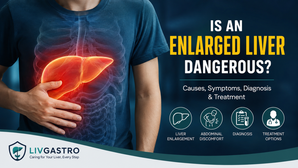
An enlarged liver, medically known as hepatomegaly, is not a disease by itself. Instead, it is usually a sign that something is affecting the liver or another part of the body. In some cases, an enlarged liver may be temporary and treatable. In others, it can indicate an underlying condition that requires timely medical attention. So, is an enlarged liver dangerous? The answer depends largely on why the liver has become enlarged, how long the enlargement has been present, and whether there is associated liver damage. Conditions ranging from fatty liver and viral hepatitis to alcohol-related liver disease, heart problems and, less commonly, tumors can cause hepatomegaly. If an ultrasound or other scan has shown that your liver is enlarged, it is important not to panic—but it should not simply be ignored either. What Does an Enlarged Liver Mean? The liver is a vital organ located primarily in the upper-right part of the abdomen. It performs numerous functions, including processing nutrients, helping regulate metabolism, producing important proteins and helping remove waste products from the body. When the liver becomes larger than expected, the condition is called hepatomegaly. An enlarged liver can develop because of inflammation, excess fat accumulation, congestion of blood within the liver, certain infections, or abnormal growths. The size of a normal liver can also vary depending on factors such as age, sex and body size, so an imaging report should always be interpreted in the appropriate clinical context. Importantly, liver enlargement does not automatically mean liver failure or cancer. The underlying cause needs to be identified before the seriousness of the condition can be determined. Is an Enlarged Liver Dangerous? An enlarged liver can be concerning, but its seriousness varies considerably from person to person. For example, liver enlargement caused by a manageable condition may improve once the underlying problem is addressed. On the other hand, hepatomegaly associated with progressive liver disease may indicate ongoing inflammation or damage that could eventually lead to complications. Some chronic liver conditions can progress to fibrosis and cirrhosis, while advanced liver disease can impair the liver’s ability to perform its essential functions. Therefore, the more useful question is not simply “Is an enlarged liver dangerous?”, but rather: “What is causing my liver to be enlarged, and is there any evidence of liver damage?” That is why medical evaluation is important after hepatomegaly is detected. What Causes an Enlarged Liver? There are several possible causes of an enlarged liver. Some are relatively common, while others require more extensive investigation. 1. Fatty Liver Disease One of the common reasons for liver enlargement is the accumulation of excess fat within liver cells. Metabolic dysfunction-associated steatotic liver disease (MASLD), previously called non-alcoholic fatty liver disease (NAFLD), can cause the liver to become enlarged. In some individuals, fatty liver can progress to metabolic dysfunction-associated steatohepatitis (MASH), where inflammation and liver injury occur. Persistent liver injury may eventually contribute to fibrosis and cirrhosis. Alcohol-Related Liver Disease Regular or excessive alcohol consumption can damage liver cells and cause inflammation. Alcohol-related hepatitis and other alcohol-associated liver conditions may result in an enlarged liver. The degree of risk depends on factors such as the amount and duration of alcohol exposure and individual susceptibility. 3. Viral Hepatitis Hepatitis viruses, including hepatitis A, B and C, can cause inflammation of the liver. Depending on the type and duration of infection, hepatitis can lead to liver enlargement and, in chronic cases, progressive liver damage. Heart-Related Conditions Liver enlargement is not always caused by a primary liver disorder. Certain heart conditions can cause blood to back up in the veins connected to the liver, resulting in congestion and enlargement. This is one reason doctors consider a person’s overall health rather than examining the liver in isolation. 5. Liver Cysts or Tumors Benign liver cysts and noncancerous growths can sometimes affect the appearance or size of the liver. In less common situations, liver cancer or cancer that has spread to the liver may also be associated with hepatomegaly. Certain Genetic or Metabolic Disorders Conditions such as hemochromatosis and Wilson’s disease can cause abnormal substances to accumulate in the liver and potentially result in liver enlargement. These conditions are less common but may require specialist evaluation. What Are the Symptoms of an Enlarged Liver? Interestingly, an enlarged liver may cause no noticeable symptoms at all. It may be discovered incidentally during a routine abdominal ultrasound or physical examination. When symptoms do occur, they can include: Discomfort or pain in the upper-right abdomen A feeling of fullness or bloating Fatigue Nausea or vomiting Reduced appetite Jaundice, causing yellowing of the skin or eyes The symptoms can vary depending on the underlying cause and whether there is associated liver dysfunction.Symptoms such as persistent abdominal swelling, significant weakness, jaundice, confusion or severe abdominal pain deserve prompt medical assessment. How Is an Enlarged Liver Diagnosed? Finding the cause of hepatomegaly generally involves more than simply measuring liver size. A gastroenterologist or hepatologist may begin with your medical history and a physical examination. After the initial evaluation, your doctor may recommend further tests to determine the underlying cause of liver enlargement. Blood Tests Liver blood tests can help assess liver injury and function. Doctors may evaluate enzymes such as ALT, AST, ALP and GGT, along with other blood parameters depending on the suspected cause. Tests for viral hepatitis or specific metabolic conditions may also be recommended. Ultrasound Doctors often recommend an abdominal ultrasound to examine the liver’s size, structure, and overall appearance. It can provide information about liver size, texture and the presence of abnormalities such as fatty changes or certain focal lesions. CT or MRI When more detailed imaging is necessary, CT or MRI may be recommended. These tests can provide additional information about the liver and surrounding structures. FibroScan or Elastography Depending on the clinical situation, non-invasive tests that assess liver stiffness may help determine whether fibrosis or scarring is present. Liver Biopsy A liver biopsy is not necessary for every person with an enlarged liver. However, in
Is Grade 2 Fatty Liver Dangerous? Understanding the Risks, Symptoms and Treatment
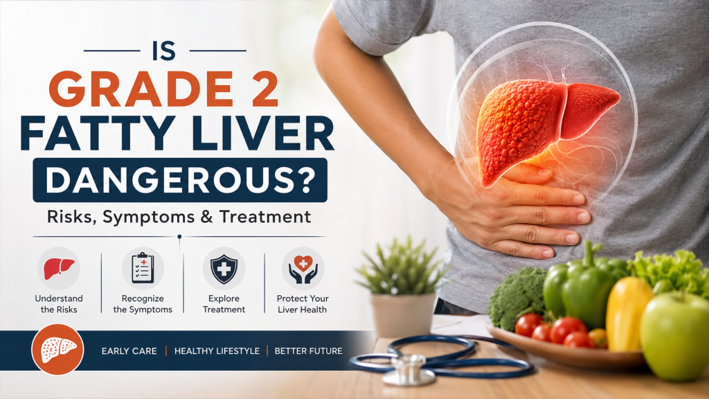
Is Grade 2 Fatty Liver Dangerous? Understanding the Risks, Symptoms and Treatment Is grade 2 fatty liver dangerous? This is a common concern after an ultrasound or liver scan reports moderate fatty infiltration of the liver. The good news is that grade 2 fatty liver does not automatically mean that your liver is permanently damaged or that you have cirrhosis. However, it should not be ignored. Grade 2 fatty liver generally indicates a moderate amount of fat accumulation in the liver. The more important question is whether there is associated liver inflammation or fibrosis (scarring). Fat accumulation, inflammation and fibrosis are different aspects of fatty liver disease, and assessing fibrosis is particularly important when determining the long-term risk. With appropriate medical evaluation and lifestyle changes, many people with fatty liver can significantly improve their liver health. What Is Grade 2 Fatty Liver? Fatty liver disease occurs when excessive fat accumulates inside liver cells. Today, metabolic dysfunction-associated steatotic liver disease (MASLD) is commonly used for fatty liver associated with metabolic risk factors such as obesity, insulin resistance, type 2 diabetes and abnormal blood lipids. When an ultrasound describes fatty liver as grade 2, it generally means a moderate degree of fatty infiltration. In some clinical grading systems, grade 2 steatosis corresponds to approximately 33–66% of liver cells containing fat, although ultrasound grading and histological grading are not identical and should not be treated as interchangeable. Importantly, grade 2 refers primarily to the amount of fat, not necessarily the amount of liver scarring. This distinction matters because a person can have substantial liver fat without advanced fibrosis, while another person with less obvious fatty liver may already have significant fibrosis. Is Grade 2 Fatty Liver Dangerous? Grade 2 fatty liver can become serious if the underlying causes are not addressed, but it is not the same as cirrhosis. Many people with fatty liver have few or no symptoms. Although many people have no noticeable symptoms, some may experience nonspecific symptoms such as fatigue or abdominal discomfort. Because fatty liver can remain silent, feeling well does not necessarily mean that the liver is completely healthy. The concern arises when fatty liver is accompanied by ongoing inflammation and liver-cell injury. This condition, historically called NASH and now commonly referred to as MASH, can contribute to fibrosis. Progressive fibrosis can eventually lead to cirrhosis and other serious complications. Therefore, the answer to “Is grade 2 fatty liver dangerous?” is: It can be a warning sign that deserves medical attention, but grade 2 fatty liver itself does not mean that you have irreversible liver damage. The next step is to determine whether there are additional risk factors and whether fibrosis is present. What Causes Grade 2 Fatty Liver? Fatty liver is frequently associated with metabolic health problems. Common contributing factors include: Overweight or obesity Insulin resistance Type 2 diabetes High triglycerides or abnormal cholesterol High blood pressure and metabolic syndrome A diet high in excess calories, refined carbohydrates or sugary foods Physical inactivity Certain medications or medical conditions Alcohol consumption, depending on the underlying diagnosis MASLD is particularly associated with cardiometabolic risk factors. This is why treating fatty liver is not only about the liver itself. Blood sugar, cholesterol, body weight, blood pressure, diet and physical activity can all be relevant to the overall management plan. What Are the Symptoms of Grade 2 Fatty Liver? One of the biggest challenges with fatty liver disease is that symptoms may be absent. Some people may experience: Fatigue or low energy A feeling of discomfort or heaviness in the upper-right abdomen Abnormal liver enzyme results on blood tests An enlarged liver in some cases However, symptoms alone cannot determine how severe fatty liver disease is. NIDDK notes that fatty liver disease is often silent and may produce few or no symptoms. This is why an ultrasound finding of grade 2 fatty liver should be discussed with a qualified gastroenterologist or hepatologist rather than simply being ignored. Does Grade 2 Fatty Liver Mean Fibrosis? Not necessarily. This is one of the most important points to understand. Steatosis means fat accumulation. Fibrosis means scar tissue formation. They are related but different processes. A report showing grade 2 fatty liver does not, by itself, tell you the stage of fibrosis. Doctors may therefore use blood-based calculations and non-invasive tests to assess the likelihood of significant liver scarring. AASLD currently provides guidance on both blood-based and imaging-based non-invasive assessment of hepatic fibrosis and steatosis. Depending on your medical history and test results, your doctor may consider: Liver function tests Complete blood count Blood glucose and HbA1c Lipid profile FIB-4 or other fibrosis risk assessment Ultrasound FibroScan or transient elastography Other specialized imaging Occasionally, liver biopsy when clinically necessary The objective is not simply to measure how much fat is present. The key clinical question is whether the liver has developed significant inflammation or fibrosis. Does Grade 2 Fatty Liver Increase the Risk of Cirrhosis? It can, but not everyone with grade 2 fatty liver develops cirrhosis. The progression generally involves a spectrum rather than an immediate jump from fatty liver to cirrhosis. In susceptible individuals, persistent liver injury may be associated with inflammation, fibrosis and eventually advanced fibrosis or cirrhosis. MASLD is recognized as a spectrum that can include steatosis, MASH, fibrosis, cirrhosis and, in some cases, liver cancer. Cirrhosis occurs when extensive scar tissue permanently damages the liver and interferes with its normal function. The risk of progression is influenced by several factors, including metabolic conditions and the presence and severity of fibrosis. That is why identifying risk early is so important. Can Grade 2 Fatty Liver Be Reversed? In many people, fat accumulation in the liver can improve significantly with appropriate lifestyle and medical management. If you are overweight or have obesity, gradual and sustainable weight reduction can reduce liver fat. NIDDK notes that losing at least 3% to 5% of body weight may reduce liver fat in people with overweight or obesity. Your doctor may recommend: 1. Improve Your
Acute Pancreatitis: Symptoms, Diagnosis & Treatment
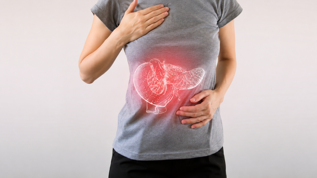
What is Acute Pancreatitis? Acute pancreatitis, also called acute pancreatic necrosis, constitutes abrupt inflammation of the pancreas that may lead to death even after receiving due medical care on account of chance development of serious complications at a later stage. However, mild types of acute pancreatitis are mostly cured through conservative treatment procedures that include total elimination of oral feeding and aggressive intravenous fluid rehydration, while severe types often involve surgery to tackle infected pancreatic necrosis and related complications. What are the symptoms of acute pancreatitis? Some of the acute pancreatitis symptoms include: ■ Severe upper abdominal pain, often radiating to the back ■ Retching ■ Shivering ■ Faster heartbeat ■ Respiratory problem ■ Hemodynamic instability (shock) ■ Hiccup ■ Fever ■ Loss of appetite ■ Hemorrhagic discoloration of the flanks What causes acute pancreatitis? ■ Excessive Alcoholism ■ Untreated gallstones ■ Penetrating ulcers ■ Carcinoma of the head of the pancreas ■ Post – ERCP complications ■ Certain drugs such as diuretics, steroids, salicylates, sulfonamides ■ Infections such as Viral hepatitis, mumps, coxsackie B virus, cytomegalovirus, Mycoplasma pneumoniae ■ Radiation X-ray ■ Autoimmune pancreatitis. What are the types of acute pancreatitis? There are basically two types of acute pancreatitis, such as Mild and Severe. Mild pancreatitis involves inflammation and edema of the pancreas, while severe pancreatitis entails necrosis of the pancreas and injury to adjacent organs. However, the hallmark of an acute pancreatitis consists of the manifestation of the inflammatory response, such as the recruitment of neutrophils to the pancreas. Nevertheless, it leads to the secondary manifestations of pancreatitis: hypovolemia from capillary permeability, acute respiratory syndrome, disseminated intravascular coagulation, renal failure, cardiovascular failure, and gastrointestinal hemorrhage. How is acute pancreatitis diagnosed? Even though acute pancreatitis may be diagnosed clinically, CT evaluation becomes necessary to distinguish between mild acute pancreatitis and severe necrotic pancreatitis. However, also relevant are Blood Investigation that include Full Blood Count, Renal Function Test, liver function, serum calcium, serum amylase and lipase, arterial blood gas, and Trypsin-Selective test. Blood studies are also conducted to identify organ failure, while they provide prognostic data about the patient’s condition. Nevertheless, triple-phase abdominal Computed Tomography, along with abdominal ultrasound, is considered in many circles as the gold standard for evaluation of acute pancreatitis. However, imaging during the first 12 hours of complaint may not prove helpful as the inflammatory and necrotic process usually takes more than 48 hours to fully manifest. Incidentally, MRI or Magnetic Resonance Imaging has also been found to be a valuable tool for visualization of the pancreas, especially for pancreatic fluid collection and necrotized debris. MRI also proves effective for patients with an allergy to the CT’s contrast material and/or overall sensitivity to vascular complications, pseudoaneyrysms as also venous thrombosis. MRI, when associated with MRCP (Magnetic Resonance Cholangiopancreatography), provides additional information in terms of the etiology of acute pancreatitis, such as the existence of small biliary stones (choledocholithiasis or cholelithiasis) and duct anomalies. Meanwhile, clinical trials have established that MRCP is no less effective a diagnostic tool for acute pancreatitis with biliary etiology than Endoscopic Retrograde Cholangiopancreatography (ERCP), while being less invasive and so more in use. Along with some of the noninvasive methods of diagnosing acute (and/or chronic) pancreatitis, Endoscopic Ultrasound (EUS) is also currently viewed as one of the most sensitive methods for detecting pancreatic lesions while in close proximity to the transducer to the pancreas. What are the modes of Treatment for acute pancreatitis? The primary state of management of patients with acute pancreatitis comprises supportive care concerning the following issues: ■ Fluid replacement through aggressive hydration at the rate of 5 to 10 ml/kg/hour of isotonic crystalloid solution, unless cardiovascular, renal, or other related comorbid factors preclude such a process. However, fluid replacement needs to be reassessed at frequent intervals in the first six hours, as well as the next 24/48 hours on the basis of clinical assessment, hematocrit, and BUN values. ■ Pain Control – Severe abdominal pain control often involves the use of intravenous opiates, mostly in the form of a patient-controlled analgesic pump. While hydromorphone or fentanyl are commonly used in acute pain control, the latter is preferred by many on account of its better safety profile, particularly in renal impairment. ■ Providing rest to the pancreas – Rest to the pancreas will also relieve the pain and the disease condition if oral feeding is eliminated. Since the pancreas is designed to secrete enzymes when food goes into the stomach, nothing will happen if there is no food there. Studies reveal that more than 20% of acute pancreatitis patients report relapse of pain within 48 hours of oral feeding. However, giving intravenous fluids will prevent dehydration and related problems. ■ Nutritional support through post-pyloric enteral feeding (in which a feeding tube is endoscopically or radiographically introduced into the third portion of the duodenum). Since this form of feeding is more physiological, it prevents gut mucosal atrophy while being free from the side effects of TPN. The other benefits of post-pyloric feeding include an inverse relationship of pancreatic exocrine secretions and remoteness of nutrient delivery from the pylorus, as also lesser risk of aspiration. Postscript: Prevention is always better than cure – try to prevent acute pancreatitis by reducing your alcohol intake. However, for people who already have an attack, treatment, in general, will take the form of support for the body functions, allowing the pancreas to heal itself on its own.
Chronic Pancreatitis: Symptoms, Causes & Treatment
What is Chronic Pancreatitis? Chronic (long-term) Pancreatitis constitutes undeviating inflammation of the pancreas that impairs your system’s ability to digest food properly and regulate the blood sugar level in your body. Although acute, short-term pancreatitis often gets cured through conservative treatment, chronic pancreatitis seldom gets permanently cured. As a matter of fact, the damage caused due to a high degree of alcoholism and other causes over an extended period of time leaves so much ill effect on the organ that no line of ethical treatment can provide enduring relief. However, acute abdominal pain and secondary symptoms can be managed through medication and other means to some extent. What are the symptoms of chronic pancreatitis? Symptoms are usually absent in the initial stage, which, however, occur as outlined below as the ailment progresses. ■ Nausea and vomiting ■ Upper abdominal pain ■ Diarrhea ■ Pale, yellowish, sticky stool ■ Undue fatigue and too much thirst ■ Inexplicable weight loss However, as the disease progresses further, more alarming symptoms begin to appear that include: ■ Typical yellowish discoloration in the eyes and the skin, indicating the onset of Jaundice ■ Presence of pancreatic fluid in the abdomen ■ Internal bleeding ■ Intestinal blockage ■ Unrelenting abdominal pain What causes chronic pancreatitis? Although there could be numerous self-induced as well as inherited causes behind the onset of chronic pancreatitis in a person’s life, the commonest of all is alcohol abuse. Medical studies reveal that more than 70% of people affected by chronic pancreatitis have a long history of excessive alcohol consumption. However, some of the other causes that are also there are listed below: ■ Blockage of the pancreatic duct either by gallstones or pancreatic stones ■ Abdominal surgery ■ High calcium levels in blood (hypercalcemia), which may have been caused by an overactive parathyroid gland (hyperparathyroidism)/li> ■ High triglyceride level in blood (hypertriglyceridemia) ■ Chronic infection that remained untreated ■ Cystic fibrosis ■ Autoimmune disease ■ Excessive cigarette smoking ■ Family history of pancreatitis ■ Incredible, yet true – ERCP may lead to chronic pancreatitis. Wonder of all wonders – sometimes no cause is found at all! How is chronic pancreatitis diagnosed? Chronic Pancreatitis is not only difficult to treat but also difficult to diagnose. For instance, blood tests that normally reveal causes related to a lot of maladies prove to be of little value in diagnosing chronic pancreatitis, at least during the early stages of this deadly disease. However, blood tests do reveal the level of pancreatic enzymes in your blood. Nevertheless, some of the following tests are conducted to obtain more definite information, while imaging tests prove more helpful in diagnosing chronic pancreatitis. ■ X-rays ■ Ultrasound ■ CT scans ■ MRI scans To obtain conclusive evidence of the extent of damage caused to your pancreas, doctors (gastroenterologists) also go for an endoscopic ultrasound. During the procedure, the doctor inserts a long, flexible tube into your mouth, down through your stomach, and right up to the small intestine. The tube contains an ultrasonic probe that emits sound waves that create detailed images of your pancreas on a monitor attached to it. What are the modes of Treatment for chronic pancreatitis? To be honest about it, there isn’t much in the medical world to cure chronic pancreatitis, because the pancreas, like the liver, once damaged, cannot be recovered back to its original condition. However, medical help is available to manage the symptoms by way of medication, endoscopic therapies, and surgery. Medications that are often prescribed to manage the symptoms include: ■ Pain management medications to suppress agonizing abdominal pain ■ Supplementary digestive enzymes if your enzyme levels are found to be much too meager for digesting food properly ■ Insulin if you become diabetic ■ Steroids, if you happen to develop autoimmune pancreatitis Endoscopic treatment of chronic pancreatitis has currently drawn benefits from endoscopic procedures previously practiced for the main bile duct. While endotherapy is widely acclaimed as the most accepted alternative to surgery, cyst drainage procedure today presents the largest step forward in that non-surgical approach, regardless of whether direct (through stomach or duodenum) or indirect through the papilla in the duodenum. Pancreatic duct drainage, associated with stone clearance, is also feasible, while the procedure gives similar results to surgery with lower morbidity. Surgery, though not indicated for all, becomes inevitable for people who cannot endure excruciating abdominal pain that does not respond to pain-killing drugs. The only alternative then entails surgical removal of part of the organ. Postscript: People suffering from chronic pancreatitis should stay away from alcohol, even if hooch hasn’t been responsible for their dreaded illness, because it will exacerbate their sickness to a great extent. They should also refrain from smoking, as it would increase their risk of developing pancreatic cancer.
Liver Cirrhosis: Symptoms, Causes & Treatment

What is Cirrhosis Liver cirrhosis, or cirrhosis, denotes a disease condition where the liver fails to function due to extensive damage over a long period of time, involving loss of liver cells, as well as irreversible scarring of the liver. Even though at the initial stage, symptoms are negligible or nil, manifestations appear in rapid succession as the disease progresses. These include fluid buildup in the abdomen, spider-like blood vessels on the skin, swelling in lower legs, and many more that are being discussed in detail next, while the patient becomes weak, loses appetite, and develops yellowish skin (Jaundice). Likely to prove fatal if not treated early. What are the symptoms of Cirrhosis? Cirrhosis symptoms may be grouped under two heads: symptoms that have resulted from failure of the liver cells and those that have occurred due to portal hypertension (secondary). Manifestations caused by the failure of the liver cells: ■ Spider angiomata or spider nevi are vascular lesions comprising a central arteriole, surrounded by many smaller vessels, occurring due to an increase in estradiol. (The formation suggests the title). ■ Palmar erythema denotes the reddening of the palms at the thenar and hypothenar eminences, caused by increased estrogen. ■ Gynecomastia denotes an increase in the breast gland in males, caused by increased estradiol. However, this is neither related to increased breast fat nor can it be called cancerous. ■ Hypogonadism denotes a decrease in sex hormones, leading to loss of libido, impotence, and testicular atrophy, resulting from suppression of hypothalamic/pituitary functions. However, this is associated with cirrhosis on account of alcoholism and hemochromatosis. ■ Abnormal liver dimensions – Enlarged as well as shrunken. ■ Ascites denotes fluid accumulation in the peritoneal cavity, giving rise to flank dullness. ■ Fetor hepaticus, denoting musty breath odor, is caused by increased dimethyl sulfide. ■ Jaundice, denoting yellowish discoloration of the skin and eyes, is caused by increased bilirubin. Manifestations caused by portal hypertension Cirrhosis (liver cirrhosis) enhances resistance to blood flow and higher pressure in the portal venous system, which causes portal hypertension, the effects of which are listed below. ■ Splenomegaly denotes an enlarged spleen. 35% to 50% patients present this picture. ■ Esophageal varices result from collateral portal blood flow through vessels in the stomach and esophagus. However, when these blood vessels become enlarged (known as varices) they are prone to rupture. This often leads to copious bleeding, which can be fatal unless immediate medical aid is made available. ■ Caput medusa relates to dilated periumbilical collateral veins associated with portal hypertension. Blood from the portal veins may be moved through these veins, and eventually to the abdominal wall veins, manifesting a model that somehow resembles the head of MEDUSA, a Greek mythological monster. ■ Cruveilhier-Baumgarten Murmur denotes venous hum that can be heard in the epigastric region (through a stethoscope) caused by collateral connections forming between the portal system and the periumbilical veins (due to portal hypertension). What causes Liver Cirrhosis? Cirrhosis is mostly caused by excessive alcohol consumption, hepatitis B, hepatitis C, as also non-alcoholic fatty liver disorders. However, the latter is again associated with obesity, high blood pressure, high body fat, and diabetes. Some of the less common causes include autoimmune hepatitis, primary biliary cirrhosis, hemochromatosis, gallstones, and certain types of medications. Cirrhosis is also typified by the substitution of normal liver tissue with scar tissue. These result in loss of liver function. How is Liver Cirrhosis diagnosed? Even though the gold standard for diagnosis of cirrhosis is liver biopsy through a percutaneous, transjugular, laparoscopic, or fine-needle method, doctors currently insist on imaging as the best diagnostic approach. However, some of these approaches are outlined below. Ultrasound is customarily used in the evaluation of cirrhosis. It may show a small and nodular liver in advanced cirrhosis, along with increased echogenicity with irregularly appearing areas. Other findings suggestive of cirrhosis in imaging are an enlarged caudate lobe, widening of the liver fissures, and enlargement of the spleen. An enlarged spleen, which normally measures less than 11–12 cm in adults, is suggestive of cirrhosis with portal hypertension in the right clinical setting. Ultrasound may also screen for hepatocellular carcinoma, portal hypertension, and Budd-Chiari syndrome (by assessing flow in the hepatic vein). Other tests performed in particular circumstances include abdominal CT and liver/bile duct MRI (MRCP). Gastroscopy (endoscopic examination of the esophagus, stomach, and duodenum) is performed in patients with established cirrhosis to exclude the possibility of esophageal varices. If these are found, prophylactic local therapy may be applied (sclerotherapy or banding), and beta-blocker treatment may be commenced. What are the lines of treatment for Cirrhosis? Since no cure exists for biliary cirrhosis, treatment generally focuses on slowing down the progress of the disease, relieving symptoms as much as possible, and preventing further complications. However, researchers are continuing to explore new drugs for treating primary biliary cirrhosis. Immunosuppressant drugs, especially methotrexate (Trexall, Rheumatrex) and colchicine (Colcrys) have been extensively tried, but their efficacy is yet to be proved.
Liver Function Test: Purpose & Procedure

Liver function tests (LFTs/LFs) conducted at LivGastro primarily consist of Blood Tests that provide vital information about the condition of a patient’s liver. Some of these tests comprise Prothrombin Time (PT/INR), aPTT, albumin, Bilirubin (both direct and indirect), and a few others. Liver transaminases (AST or SGOT and ALT/SGPT ) provide helpful biomarkers in terms of the types of liver injury a patient has suffered. Thanks to the high-tech medical equipment from the manufacturer, Hisky Med, for equipping this digestive hospital in the City of Joy with state-of-the-art medical instruments! Without them, it could not have offered accurate, convenient, and standard services to the locals approaching this diagnostic centre to diagnose chronic liver diseases. As luck would have it, call it ill luck, most liver disorders cause not so intense initial symptoms, while ninety percent of hepatic disease conditions have a poor prognosis. However, tests are essential to ascertain the extent of damage caused to the person’s liver and to determine the right course of treatment. While some tests are associated with functionality, such as albumen, there are others that relate to cellular integrity, such as transaminase, while some others are linked to the biliary tract (gamma-glutamyl transferase and alkaline phosphate). Some biochemical tests are also conducted for the evaluation and management of patients with hepatic dysfunction, especially in terms of ascertaining the extent of damage caused to the liver, as well as the line of treatment that would best suit such patients. Other tests commonly conducted at LivGastro include the following: ■ 5’ Nucleotidase (5’NTD), which is specific for cholestasis that refers to damage to the intra- or extrahepatic biliary system, is often used as a substitute for GGT in ascertaining if the elevated ALP is of biliary or extrabiliary origin. ■ Coagulation Test – The liver is responsible for the production of the vast majority of coagulation factors. In patients with liver disease, INR can be used as a marker of liver synthetic function, as it includes factor VII, which has the shortest half-life (2-6 hours) of all coagulation factors measured in INR. An elevated INR in patients with liver disease, however, does not necessarily mean the patient tends to bleed, as it only measures procoagulants and not anticoagulants. In liver disease, the synthesis of both is decreased, and some patients are even found to be hypercoagulable (increased tendency to clot) despite an elevated INR. In liver patients, coagulation is better determined by more modern tests such as thromboelastogram (TEG) or thromboelastrometry (ROTEM). ■ Serum glucose Test, also called ‘BG’ or ‘Glu,’ is conducted to measure the liver’s ability to produce glucose (gluconeogenesis). In fact, it is the terminal function to be lost in the setting of ‘fulminant liver failure’. ■ Lactate dehydrogenase Test – Lactate dehydrogenase or LDH is observed in most body tissues, including the liver. However, elevated LDH levels often indicate liver damage. However, LDH isotype-1 (or cardiac) is often used for estimating damage caused to cardiac tissues, even though troponin and creatine kinase tests are found to be better alternatives.
Fatty Liver Disease: Symptoms, Diagnosis & Treatment
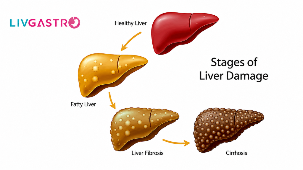
Fatty Liver, as the name suggests, is the excessive build-up of fat in the liver of about 5-10% of the liver’s weight. Most of the time, it’s not a serious concern, but in some cases, it is something to fret about, as it leads to liver damage. It starts with inflammation, causing the tissues to damage. The medical term for this first stage is steatohepatitis. In the second stage, scar tissue begins to form, which in turn damages the liver. Doctors call it fibrosis. In the third and final stage, the healthy tissues get replaced by extensive scar tissue. This is when cirrhosis of the liver occurs. It is a final blow to the liver, whereby liver functioning starts slowing down. Sooner or later, the liver function gets blocked entirely. According to the Cleveland Clinic, this cirrhosis transpires into liver cancer or liver failure in patients. Do you now get why fatty liver disease is a thing to panic about? Forms of Fatty Liver Disease Fatty liver disease has two primary forms: Alcohol-induced – As hinted, it is caused by heavy drinking. Non-alcoholic – Alternatively called NAFLD or Non-Alcoholic Fatty Liver Disease, the cause is still unknown, but diabetes and obesity have been said to increase the risk of occurrence. Symptoms to Pay Attention The symptoms of fatty liver are often silent unless the disease progresses to liver cirrhosis. The signs to watch out for are: ■ Weight loss, appetite loss, and nausea ■ Extreme weakness, exhaustion, and mental confusion ■ Jaundice ■ Abdominal pain, especially in the upper right side of the abdomen ■ Edema or swollen legs and abdomen If you ever feel any of the symptoms above, be prompt and book an appointment with a hepatologist for diagnostic services. The next section covers the Liver function tests, or a diagnosis that a doctor may order. Diagnosis Fatty liver silently creeps in, and for a layperson, it is challenging to spot the same. However, a doctor can easily determine the condition by noticing the elevation in liver enzymes from blood tests done for other ailments. To map the liver injury, a doctor often orders the tests mentioned below: ■ A liver biopsy to check the stage the liver disease has reached ■ Ultrasound or a CT Scan to examine the liver condition ■ FibroScan to determine the fat content in the liver Treatment for Fatty Liver Disease Unfortunately, there is no medication for treating fatty liver disease. Doctors primarily advise altering lifestyles to improve health conditions. Therefore, the treatment mostly comprises the following: ■ Reducing weight ■ Intake of thiazolidinediones or vitamin E ■ Avoiding alcohol consumption ■ Intake of medications to control cholesterol, diabetes, and triglycerides How to Prevent Fatty Liver Disease? If you have not been hit by fatty liver disease, you can do the following to maintain your overall health: ■ Get yourself involved in regular exercise ■ Maintain a healthy weight ■ Take prescribed medications ■ Put a check on alcohol consumption Reverse Your Fatty Liver Disease! Yes, you read it right. It can be either through a weight loss program or by avoiding alcohol. A Mediterranean diet, where one needs to consume lots of vegetables, fruits, and good fats, can lead to steady weight loss. So, if you are living with the ailment, don’t worry! Just make sure it never progresses into liver cirrhosis, and even if it does, go for immediate treatment to prevent liver failure or cancer.
Capsule Endoscopy: What Gastroenterologists Want You to Know
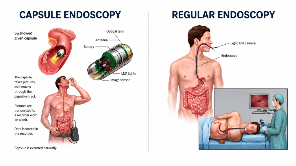
You have probably had a colonoscopy recommended, or perhaps an upper endoscopy. What about the stretch of digestive tract in between – the small intestine – that stretches nearly six metres and sits largely out of reach of conventional scopes? That’s where capsule endoscopy comes in. If you have been dealing with persistent abdominal pain, unexplained bleeding, or recurring digestive discomfort and have not found answers yet, consulting the best gastroenterologist you can find might lead you straight to this procedure. It’s less intimidating than it sounds, and it often reveals what everything else misses. What is Capsule Endoscopy and How Does It Work? Capsule endoscopy is a diagnostic procedure in which you swallow a small capsule — roughly the size of a large vitamin tablet — containing a tiny camera, a light source, and a radio transmitter. Once swallowed, the capsule travels naturally through your digestive tract, from the oesophagus down to the small intestine, capturing around 10,000 photographs along the way. These images are transmitted wirelessly to a small receiver worn around your waist. The whole process takes about eight hours, after which the capsule passes out of your body through stool. Before the procedure, your doctor will administer laxatives to clear the digestive system of food debris. So the camera can capture clear images. There’s no sedation involved. You simply swallow the capsule and go about your day as usual while it does its work inside. What Conditions Can Capsule Endoscopy Detect? This is where capsule endoscopy earns its reputation. It’s particularly effective at identifying problems in the small intestine that standard endoscopy and colonoscopy cannot reach. Conditions it can help diagnose include: ■ Unexplained gastrointestinal bleeding — when upper endoscopy and colonoscopy have drawn a blank ■ Crohn’s disease — especially early-stage or small bowel involvement ■ Intestinal tumours — both benign and malignant growths ■ Angiodysplasias — abnormal blood vessels in the gut wall ■ Celiac disease — where damage to the small intestinal lining needs to be assessed ■ Colon polyps and inflammatory bowel disease (IBD) Symptoms that might prompt a referral to the best gastroenterologist for this test include abdominal cramping, blood in the stool, persistent diarrhoea, chronic fatigue linked to unexplained anaemia, or significant unintended weight loss. Who is Capsule Endoscopy For? Capsule endoscopy is recommended for a fairly specific group of patients — those for whom conventional diagnostic methods have been inconclusive or insufficient. ■ People who have been bleeding somewhere in the gut, and other tests have come up empty ■ Those with suspected or confirmed Crohn’s disease, where the small bowel needs a proper look ■ Anyone dealing with ongoing abdominal pain or weight loss that has not been explained yet ■ Patients keeping an eye on existing polyps or intestinal growths ■ Those with persistent diarrhoea or signs that the body isn’t absorbing nutrients the way it should — celiac disease being a common culprit One thing worth knowing: if you have a history of narrowing or blockages in the intestine, your doctor will want to discuss this before proceeding. There’s a small chance the capsule could get lodged, so your gastroenterologist will review your history and decide whether the procedure is right for you. How is Capsule Endoscopy Different from a Regular Endoscopy? The most obvious difference is comfort. There’s no scope insertion, no sedation, and no recovery time needed afterwards. You swallow a pill and carry on. That said, capsule endoscopy does have limitations worth understanding: The capsule cannot perform therapeutic interventions — it cannot take biopsies or remove polyps If the capsule moves too quickly, images may be blurred or incomplete If battery life runs out before the capsule completes its journey, areas may be missed Pinpointing the exact location of an abnormality can sometimes be difficult These are procedural realities, not reasons to avoid it. In many cases, capsule endoscopy remains the most practical and informative way to evaluate the small bowel — and that makes it invaluable. Book Your Capsule Endoscopy at LivGastro — Kolkata’s Trusted Gastroenterology Hospital At LivGastro, every capsule endoscopy is overseen by Dr. Vijay Kumar Rai — a gastroenterologist recognised by both the American College of Gastroenterology and the American Society of GI Endoscopy. That’s not just a line on a CV — it means the person overseeing your diagnosis is trained to international standards. We use equipment sourced from Germany, Japan, and America, and every test is carried out by qualified doctors, not left to junior staff. If you’ve been looking up “acidity doctor near me” or trying to find a gastroenterology hospital in Kolkata where you can actually trust who’s doing your tests, LivGastro is worth a visit. All diagnostic procedures at LivGastro are conducted by highly trained doctors under Dr. Rai’s supervision, and results are delivered promptly so your care can move forward without delay. We provide complete day care gastro and liver services — consultation, diagnostics, pharmacy, and nutrition — all under one roof. Reach us at +91 9147105221 or WhatsApp 9369120028 to book your appointment. We are available Monday to Saturday, 8 am to 10 pm.
Prevention of Colorectal Cancer Through Early Detection
What Are The Major Risk Factors? Some of the key risk factors that cause colorectal cancer include family history (of the disease) as well as old age, while obesity, excessive alcohol use, indolence, and cigarette smoking are not ignored either. Alongside, people with a history of inflammatory bowel disease, such as ulcerative colitis and Crohn’s disease, reportedly run a higher risk of having colorectal cancer than those without such conditions. Screening tests for early diagnosis of colorectal cancer often prevent the spread of cancer. Quite a few screening tests have since been developed to help gastroenterologists such as Dr. V.K. Rai, M.D., and others detect colorectal cancer early, when the disease becomes more treatable. What’s more, some tests that detect or identify the adenomas are known to prevent the development of cancer through proper medical intervention. So, the screening procedure may also be considered as a form of cancer prevention method in most cases. What Tests Are Recommended For Diagnosing Colorectal Cancer? The following tests and/or investigative procedures are recommended for diagnosing colorectal cancer. High-sensitivity Fecal Occult Blood Test (FOBT) – Both polyps and colorectal cancers can bleed, while FOBT checks for tiny amounts of blood in feces (stool) that cannot be seen visually. (Blood in stool may also indicate the presence of conditions that are not cancerous, such as hemorrhoids. As of now, two types of FOBT are approved by the FDA for screening colorectal cancer: guaiac FOBT (gFOBT) and the fecal immunochemical (or immunohistochemical) test (FIT or iFOBT). With both types of FOBT, stool samples collected by the patient with the help of a kit are sent to the gastroenterologist. ■ Guaiac FOBT uses a chemical to detect heme, a component of the blood protein hemoglobin. Since the guaiac FOBT can also detect heme in some foods (for example, red meat), people have to avoid certain foods before having this test. ■ FIT uses antibodies to detect human hemoglobin protein specifically (4, 5). Dietary restrictions are usually not applicable in FIT. Studies, however, have revealed that guaiac FOBT, when performed every 1 to 2 years in people aged 50 to 80 years, can help reduce the number of deaths (due to colorectal cancer) by 15 to 33%. When FOBT is the only type of colorectal cancer screening test recommended, experts usually suggest yearly testing. Stool DNA test (FIT-DNA) is the only stool DNA test that has been approved by the FDA, while Cologuard® is a multitarget test that detects tiny amounts of blood in stool (with an immunochemical test similar to FIT) as well as nine DNA biomarkers in three genes that have been found in colorectal cancer and precancerous advanced adenomas. The DNA comes from cells in the lining of the colon and rectum that are shed and collected in stool as it passes through the large intestine and rectum. As with both types of FOBT, the stool sample for the FIT-DNA test is collected by the patient using a kit, and the sample is mailed to a laboratory for testing. A computer program analyzes the results of the two tests (blood and DNA biomarkers) and provides a finding of negative or positive. People who have a positive finding with this test are advised to have a colonoscopy. In one study comprising people who were at average risk of developing colon cancer but had no symptoms of colon problems, this test detected more cancers and adenomas than the FIT test (that is, it was more sensitive). However, the FIT-DNA test was also more likely to identify an abnormality when none was actually present (that is, it had more false-positive results). Sigmoidoscopy In this test, the rectum and sigmoid colon are examined using a sigmoidoscope, which is a flexible lighted tube with a lens for viewing and a tool for removing tissue. This instrument is inserted through the anus into the rectum and sigmoid colon as air (or carbon dioxide) is pumped into the colon to expand it so the gastroenterologist may be able to view the colon lining more effectively. During sigmoidoscopy, abnormal growths in the rectum and sigmoid colon can be removed for analysis (biopsied). The lower colon must be cleared of stool before sigmoidoscopy, but the preparation is less extensive than that required for colonoscopy. People are usually not sedated for this test. Studies, however, have shown that people who have regular screening with sigmoidoscopy after age 50 years have a 60 to 70% lower risk of death due to cancer of the rectum and lower colon than people who do not have screening. One randomized controlled clinical trial found that even just one sigmoidoscopy screening between the ages of 55 and 64 can substantially reduce colorectal cancer incidence as well as mortality. Experts generally recommend sigmoidoscopy every 5 years with or without gFOBT or FIT every 3 years for people at average risk who have had negative test results. Standard [Optical] Colonoscopy In this test, the rectum and entire colon are examined by using a colonoscope, which is a flexible lighted tube with a lens for viewing and a tool for removing tissue. Like the shorter sigmoidoscope, the colonoscope is inserted through the anus into the rectum and the colon, as air (or carbon dioxide) is pumped into the colon to expand it so the doctor can see the colon lining more clearly. During a colonoscopy, any abnormal growths in the colon and the rectum can be removed, including growths in the upper parts of the colon that are not reached by sigmoidoscopy. However, a thorough cleansing of the entire colon is necessary before this test. Most patients receive some form of sedation during the test. Studies reveal that colonoscopy reduces deaths from colorectal cancer by about 60 to 70%. Additional studies are currently being done to better evaluate how effective colonoscopy screening methods are. Experts recommend a colonoscopy every 10 years for people at average risk as long as their test results are negative. Virtual Colonoscopy This screening method, often called computed