
Liver Cirrhosis: Symptoms, Causes & Treatment

What is Cirrhosis Liver cirrhosis, or cirrhosis, denotes a disease condition where the liver fails to function due to extensive damage over a long period of time, involving loss of liver cells, as well as irreversible scarring of the liver. Even though at the initial stage, symptoms are negligible or nil, manifestations appear in rapid succession as the disease progresses. These include fluid buildup in the abdomen, spider-like blood vessels on the skin, swelling in lower legs, and many more that are being discussed in detail next, while the patient becomes weak, loses appetite, and develops yellowish skin (Jaundice). Likely to prove fatal if not treated early. What are the symptoms of Cirrhosis? Cirrhosis symptoms may be grouped under two heads: symptoms that have resulted from failure of the liver cells and those that have occurred due to portal hypertension (secondary). Manifestations caused by the failure of the liver cells: ■ Spider angiomata or spider nevi are vascular lesions comprising a central arteriole, surrounded by many smaller vessels, occurring due to an increase in estradiol. (The formation suggests the title). ■ Palmar erythema denotes the reddening of the palms at the thenar and hypothenar eminences, caused by increased estrogen. ■ Gynecomastia denotes an increase in the breast gland in males, caused by increased estradiol. However, this is neither related to increased breast fat nor can it be called cancerous. ■ Hypogonadism denotes a decrease in sex hormones, leading to loss of libido, impotence, and testicular atrophy, resulting from suppression of hypothalamic/pituitary functions. However, this is associated with cirrhosis on account of alcoholism and hemochromatosis. ■ Abnormal liver dimensions – Enlarged as well as shrunken. ■ Ascites denotes fluid accumulation in the peritoneal cavity, giving rise to flank dullness. ■ Fetor hepaticus, denoting musty breath odor, is caused by increased dimethyl sulfide. ■ Jaundice, denoting yellowish discoloration of the skin and eyes, is caused by increased bilirubin. Manifestations caused by portal hypertension Cirrhosis (liver cirrhosis) enhances resistance to blood flow and higher pressure in the portal venous system, which causes portal hypertension, the effects of which are listed below. ■ Splenomegaly denotes an enlarged spleen. 35% to 50% patients present this picture. ■ Esophageal varices result from collateral portal blood flow through vessels in the stomach and esophagus. However, when these blood vessels become enlarged (known as varices) they are prone to rupture. This often leads to copious bleeding, which can be fatal unless immediate medical aid is made available. ■ Caput medusa relates to dilated periumbilical collateral veins associated with portal hypertension. Blood from the portal veins may be moved through these veins, and eventually to the abdominal wall veins, manifesting a model that somehow resembles the head of MEDUSA, a Greek mythological monster. ■ Cruveilhier-Baumgarten Murmur denotes venous hum that can be heard in the epigastric region (through a stethoscope) caused by collateral connections forming between the portal system and the periumbilical veins (due to portal hypertension). What causes Liver Cirrhosis? Cirrhosis is mostly caused by excessive alcohol consumption, hepatitis B, hepatitis C, as also non-alcoholic fatty liver disorders. However, the latter is again associated with obesity, high blood pressure, high body fat, and diabetes. Some of the less common causes include autoimmune hepatitis, primary biliary cirrhosis, hemochromatosis, gallstones, and certain types of medications. Cirrhosis is also typified by the substitution of normal liver tissue with scar tissue. These result in loss of liver function. How is Liver Cirrhosis diagnosed? Even though the gold standard for diagnosis of cirrhosis is liver biopsy through a percutaneous, transjugular, laparoscopic, or fine-needle method, doctors currently insist on imaging as the best diagnostic approach. However, some of these approaches are outlined below. Ultrasound is customarily used in the evaluation of cirrhosis. It may show a small and nodular liver in advanced cirrhosis, along with increased echogenicity with irregularly appearing areas. Other findings suggestive of cirrhosis in imaging are an enlarged caudate lobe, widening of the liver fissures, and enlargement of the spleen. An enlarged spleen, which normally measures less than 11–12 cm in adults, is suggestive of cirrhosis with portal hypertension in the right clinical setting. Ultrasound may also screen for hepatocellular carcinoma, portal hypertension, and Budd-Chiari syndrome (by assessing flow in the hepatic vein). Other tests performed in particular circumstances include abdominal CT and liver/bile duct MRI (MRCP). Gastroscopy (endoscopic examination of the esophagus, stomach, and duodenum) is performed in patients with established cirrhosis to exclude the possibility of esophageal varices. If these are found, prophylactic local therapy may be applied (sclerotherapy or banding), and beta-blocker treatment may be commenced. What are the lines of treatment for Cirrhosis? Since no cure exists for biliary cirrhosis, treatment generally focuses on slowing down the progress of the disease, relieving symptoms as much as possible, and preventing further complications. However, researchers are continuing to explore new drugs for treating primary biliary cirrhosis. Immunosuppressant drugs, especially methotrexate (Trexall, Rheumatrex) and colchicine (Colcrys) have been extensively tried, but their efficacy is yet to be proved.
Fatty Liver Disease: Symptoms, Diagnosis & Treatment
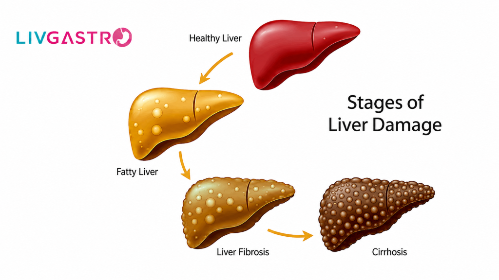
Fatty Liver, as the name suggests, is the excessive build-up of fat in the liver of about 5-10% of the liver’s weight. Most of the time, it’s not a serious concern, but in some cases, it is something to fret about, as it leads to liver damage. It starts with inflammation, causing the tissues to damage. The medical term for this first stage is steatohepatitis. In the second stage, scar tissue begins to form, which in turn damages the liver. Doctors call it fibrosis. In the third and final stage, the healthy tissues get replaced by extensive scar tissue. This is when cirrhosis of the liver occurs. It is a final blow to the liver, whereby liver functioning starts slowing down. Sooner or later, the liver function gets blocked entirely. According to the Cleveland Clinic, this cirrhosis transpires into liver cancer or liver failure in patients. Do you now get why fatty liver disease is a thing to panic about? Forms of Fatty Liver Disease Fatty liver disease has two primary forms: Alcohol-induced – As hinted, it is caused by heavy drinking. Non-alcoholic – Alternatively called NAFLD or Non-Alcoholic Fatty Liver Disease, the cause is still unknown, but diabetes and obesity have been said to increase the risk of occurrence. Symptoms to Pay Attention The symptoms of fatty liver are often silent unless the disease progresses to liver cirrhosis. The signs to watch out for are: ■ Weight loss, appetite loss, and nausea ■ Extreme weakness, exhaustion, and mental confusion ■ Jaundice ■ Abdominal pain, especially in the upper right side of the abdomen ■ Edema or swollen legs and abdomen If you ever feel any of the symptoms above, be prompt and book an appointment with a hepatologist for diagnostic services. The next section covers the Liver function tests, or a diagnosis that a doctor may order. Diagnosis Fatty liver silently creeps in, and for a layperson, it is challenging to spot the same. However, a doctor can easily determine the condition by noticing the elevation in liver enzymes from blood tests done for other ailments. To map the liver injury, a doctor often orders the tests mentioned below: ■ A liver biopsy to check the stage the liver disease has reached ■ Ultrasound or a CT Scan to examine the liver condition ■ FibroScan to determine the fat content in the liver Treatment for Fatty Liver Disease Unfortunately, there is no medication for treating fatty liver disease. Doctors primarily advise altering lifestyles to improve health conditions. Therefore, the treatment mostly comprises the following: ■ Reducing weight ■ Intake of thiazolidinediones or vitamin E ■ Avoiding alcohol consumption ■ Intake of medications to control cholesterol, diabetes, and triglycerides How to Prevent Fatty Liver Disease? If you have not been hit by fatty liver disease, you can do the following to maintain your overall health: ■ Get yourself involved in regular exercise ■ Maintain a healthy weight ■ Take prescribed medications ■ Put a check on alcohol consumption Reverse Your Fatty Liver Disease! Yes, you read it right. It can be either through a weight loss program or by avoiding alcohol. A Mediterranean diet, where one needs to consume lots of vegetables, fruits, and good fats, can lead to steady weight loss. So, if you are living with the ailment, don’t worry! Just make sure it never progresses into liver cirrhosis, and even if it does, go for immediate treatment to prevent liver failure or cancer.
Chronic Liver Disease ICD-10: Complete Coding Guide for Clinicians & Coders
Accurate coding of chronic liver disease ICD-10 is one of the most critical — and frequently mishandled — tasks in medical billing and clinical documentation. With over 1.5 billion people worldwide affected by chronic liver conditions, getting the ICD-10 codes right impacts reimbursement, outcomes tracking, and care quality. This guide covers everything from the correct code structure to common pitfalls coders face daily. What Is Chronic Liver Disease? Chronic liver disease (CLD) refers to a progressive deterioration of liver function lasting six months or longer. Unlike acute liver conditions, CLD unfolds gradually — damaging liver cells, disrupting metabolic processes, and ultimately impairing the organ’s ability to filter blood, produce proteins, and metabolize nutrients. The condition spans a wide spectrum: from early-stage non-alcoholic fatty liver disease (NAFLD) and alcoholic hepatitis to the advanced stages of fibrosis, cirrhosis, and hepatocellular carcinoma. Because CLD rarely presents with dramatic early symptoms, it often goes undetected until significant organ damage has occurred. Understanding the ICD-10-CM Code Structure for Chronic Liver Disease The ICD-10-CM (International Classification of Diseases, 10th Revision, Clinical Modification) provides a hierarchical coding system for liver diseases under Chapter 11: Diseases of the Digestive System (K00–K95). Chronic liver disease codes primarily fall under the K70–K77 block — “Diseases of the Liver.” Understanding how these codes are organized is the first step toward accurate documentation. Primary Chronic Liver Disease ICD-10 Codes at a Glance ICD-10 Code Description Key Notes K70.30 Alcoholic cirrhosis of liver without ascites Specify alcohol use disorder separately K70.31 Alcoholic cirrhosis of liver with ascites Common in advanced alcoholic CLD K70.10 Alcoholic hepatitis without ascites Distinguish from acute episode K70.11 Alcoholic hepatitis with ascites High mortality risk; document carefully K73.0 Chronic persistent hepatitis, NEC Use when type unspecified K73.1 Chronic lobular hepatitis, NEC Histological diagnosis required K73.2 Chronic active hepatitis, NEC Also called chronic aggressive hepatitis K73.9 Chronic hepatitis, unspecified Use only when type cannot be determined K74.0 Hepatic fibrosis Code also underlying cause K74.1 Hepatic sclerosis Rare; usually secondary to other liver disease K74.60 Unspecified cirrhosis of liver Use when etiology undocumented K74.69 Other cirrhosis of liver Includes cryptogenic cirrhosis K76.0 Fatty (change of) liver, NEC Used for NAFLD without inflammation K75.81 Nonalcoholic steatohepatitis (NASH) NASH — important metabolic liver disease code K76.89 Other specified diseases of liver Covers conditions not elsewhere classified Coder’s Pro Tip:- Always document the etiology of liver disease (alcoholic, viral, metabolic, autoimmune) before defaulting to an “unspecified” code. Specificity drives reimbursement accuracy and helps flag high-risk patients in population health programs. Chronic Liver Disease: Stages and Their ICD-10 Implications Chronic liver disease progresses through distinct stages. Each stage has different clinical, functional, and coding implications. Coders and clinicians must understand disease staging to select the most appropriate and specific ICD-10 code. Hepatic Inflammation — The earliest stage involves inflammation of liver cells (hepatocytes). Codes such as K73.2 (Chronic active hepatitis) or K75.81 (NASH) apply at this stage. Biopsy or imaging findings typically confirm the diagnosis. Hepatic Fibrosis (K74.0) — Repeated injury triggers scar tissue formation. Fibrosis does not present with obvious clinical symptoms but is confirmed via FibroScan, FIB-4 score, or biopsy. This is a critical transitional code to capture before cirrhosis develops. Compensated Cirrhosis — The liver still performs most functions despite significant scarring. ICD-10 codes K74.60 or K70.30 apply depending on etiology. These patients may appear clinically stable yet face high long-term risk. Decompensated Cirrhosis — Characterized by complications: ascites, variceal bleeding, hepatic encephalopathy, jaundice. Ascites specificity (e.g., K70.31) is mandatory here. Additional complication codes should be sequenced correctly. End-Stage Liver Disease (ESLD) / Hepatocellular Carcinoma — This stage may require codes from the neoplasm chapter (C22.0 for hepatocellular carcinoma) along with the underlying chronic liver disease code. Liver transplant evaluation codes may also apply. Signs and Symptoms That Should Trigger CLD Workup Recognizing early symptoms supports both clinical care and timely, accurate coding. Many CLD patients present with overlapping, nonspecific complaints — making documentation and specificity even more important. Jaundice Yellowing of skin and eyes; signals bilirubin buildup Ascites Abdominal fluid accumulation; document with/without in code Hepatic Encephalopathy Confusion and cognitive changes due to toxin buildup Esophageal Varices Portal hypertension causes dilated esophageal veins Fatigue & Weakness Early nonspecific symptom; flag for further workup Spider Angiomata Dilated skin vessels; clinical sign of CLD Common Coding Errors in Chronic Liver Disease ICD-10 Medical coding for chronic liver disease is error-prone — especially in busy outpatient settings. Below are the most frequent mistakes coders and physicians make: Using K74.60 (unspecified cirrhosis) when a specific etiology such as alcohol or NASH has been clearly documented. Failing to code ascites specificity — the “with ascites” subcode dramatically affects DRG assignment and risk-scoring. Omitting alcohol use disorder codes (F10.10–F10.29) when coding alcoholic liver disease — these are required additional codes per ICD-10-CM guidelines. Sequencing errors: the underlying liver disease should generally be listed as the principal diagnosis, not the complication. Confusing K75.81 (NASH) with K76.0 (fatty liver/NAFLD without steatohepatitis) — these are distinct conditions with different clinical severity. Missing hepatitis virus codes (B18.0–B18.9) as required additional codes when chronic viral hepatitis underlies the CLD. Documentation Best Practice:- Physicians should document the following in every CLD encounter: etiology of liver disease, presence or absence of ascites, current stage (fibrosis vs. cirrhosis), history of decompensation events, and substance use history. This directly enables specific, defensible ICD-10 code selection. ICD-10 Code Description B18.0 Chronic viral hepatitis B with delta-agent B18.1 Chronic viral hepatitis B without delta-agent B18.2 Chronic viral hepatitis C B18.8 Other chronic viral hepatitis B18.9 Chronic viral hepatitis, unspecified ICD-10 Coding for Chronic Liver Disease Complications Advanced CLD generates a cascade of complications — each requiring its own accurate code in addition to the primary liver disease code. Capturing these codes completely is essential for correct risk adjustment and quality measure performance. K76.6 — Portal hypertension I85.00 / I85.01 — Esophageal varices without / with bleeding K72.10 / K72.11 — Chronic hepatic failure without / with coma R18.0 — Malignant ascites (when ascites is due to malignancy) K76.7 — Hepatorenal syndrome G93.41 — Metabolic encephalopathy (hepatic encephalopathy)