
Chronic Pancreatitis: Symptoms, Causes & Treatment
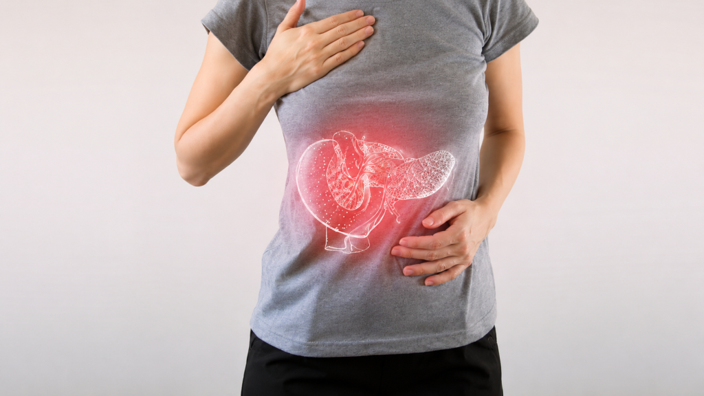
What is Chronic Pancreatitis? Chronic (long-term) Pancreatitis constitutes undeviating inflammation of the pancreas that impairs your system’s ability to digest food properly and regulate the blood sugar level in your body. Although acute, short-term pancreatitis often gets cured through conservative treatment, chronic pancreatitis seldom gets permanently cured. As a matter of fact, the damage caused due to a high degree of alcoholism and other causes over an extended period of time leaves so much ill effect on the organ that no line of ethical treatment can provide enduring relief. However, acute abdominal pain and secondary symptoms can be managed through medication and other means to some extent. What are the symptoms of chronic pancreatitis? Symptoms are usually absent in the initial stage, which, however, occur as outlined below as the ailment progresses. ■ Nausea and vomiting ■ Upper abdominal pain ■ Diarrhea ■ Pale, yellowish, sticky stool ■ Undue fatigue and too much thirst ■ Inexplicable weight loss However, as the disease progresses further, more alarming symptoms begin to appear that include: ■ Typical yellowish discoloration in the eyes and the skin, indicating the onset of Jaundice ■ Presence of pancreatic fluid in the abdomen ■ Internal bleeding ■ Intestinal blockage ■ Unrelenting abdominal pain What causes chronic pancreatitis? Although there could be numerous self-induced as well as inherited causes behind the onset of chronic pancreatitis in a person’s life, the commonest of all is alcohol abuse. Medical studies reveal that more than 70% of people affected by chronic pancreatitis have a long history of excessive alcohol consumption. However, some of the other causes that are also there are listed below: ■ Blockage of the pancreatic duct either by gallstones or pancreatic stones ■ Abdominal surgery ■ High calcium levels in blood (hypercalcemia), which may have been caused by an overactive parathyroid gland (hyperparathyroidism)/li> ■ High triglyceride level in blood (hypertriglyceridemia) ■ Chronic infection that remained untreated ■ Cystic fibrosis ■ Autoimmune disease ■ Excessive cigarette smoking ■ Family history of pancreatitis ■ Incredible, yet true – ERCP may lead to chronic pancreatitis. Wonder of all wonders – sometimes no cause is found at all! How is chronic pancreatitis diagnosed? Chronic Pancreatitis is not only difficult to treat but also difficult to diagnose. For instance, blood tests that normally reveal causes related to a lot of maladies prove to be of little value in diagnosing chronic pancreatitis, at least during the early stages of this deadly disease. However, blood tests do reveal the level of pancreatic enzymes in your blood. Nevertheless, some of the following tests are conducted to obtain more definite information, while imaging tests prove more helpful in diagnosing chronic pancreatitis. ■ X-rays ■ Ultrasound ■ CT scans ■ MRI scans To obtain conclusive evidence of the extent of damage caused to your pancreas, doctors (gastroenterologists) also go for an endoscopic ultrasound. During the procedure, the doctor inserts a long, flexible tube into your mouth, down through your stomach, and right up to the small intestine. The tube contains an ultrasonic probe that emits sound waves that create detailed images of your pancreas on a monitor attached to it. What are the modes of Treatment for chronic pancreatitis? To be honest about it, there isn’t much in the medical world to cure chronic pancreatitis, because the pancreas, like the liver, once damaged, cannot be recovered back to its original condition. However, medical help is available to manage the symptoms by way of medication, endoscopic therapies, and surgery. Medications that are often prescribed to manage the symptoms include: ■ Pain management medications to suppress agonizing abdominal pain ■ Supplementary digestive enzymes if your enzyme levels are found to be much too meager for digesting food properly ■ Insulin if you become diabetic ■ Steroids, if you happen to develop autoimmune pancreatitis Endoscopic treatment of chronic pancreatitis has currently drawn benefits from endoscopic procedures previously practiced for the main bile duct. While endotherapy is widely acclaimed as the most accepted alternative to surgery, cyst drainage procedure today presents the largest step forward in that non-surgical approach, regardless of whether direct (through stomach or duodenum) or indirect through the papilla in the duodenum. Pancreatic duct drainage, associated with stone clearance, is also feasible, while the procedure gives similar results to surgery with lower morbidity. Surgery, though not indicated for all, becomes inevitable for people who cannot endure excruciating abdominal pain that does not respond to pain-killing drugs. The only alternative then entails surgical removal of part of the organ. Postscript: People suffering from chronic pancreatitis should stay away from alcohol, even if hooch hasn’t been responsible for their dreaded illness, because it will exacerbate their sickness to a great extent. They should also refrain from smoking, as it would increase their risk of developing pancreatic cancer.
Liver Cirrhosis: Symptoms, Causes & Treatment

What is Cirrhosis Liver cirrhosis, or cirrhosis, denotes a disease condition where the liver fails to function due to extensive damage over a long period of time, involving loss of liver cells, as well as irreversible scarring of the liver. Even though at the initial stage, symptoms are negligible or nil, manifestations appear in rapid succession as the disease progresses. These include fluid buildup in the abdomen, spider-like blood vessels on the skin, swelling in lower legs, and many more that are being discussed in detail next, while the patient becomes weak, loses appetite, and develops yellowish skin (Jaundice). Likely to prove fatal if not treated early. What are the symptoms of Cirrhosis? Cirrhosis symptoms may be grouped under two heads: symptoms that have resulted from failure of the liver cells and those that have occurred due to portal hypertension (secondary). Manifestations caused by the failure of the liver cells: ■ Spider angiomata or spider nevi are vascular lesions comprising a central arteriole, surrounded by many smaller vessels, occurring due to an increase in estradiol. (The formation suggests the title). ■ Palmar erythema denotes the reddening of the palms at the thenar and hypothenar eminences, caused by increased estrogen. ■ Gynecomastia denotes an increase in the breast gland in males, caused by increased estradiol. However, this is neither related to increased breast fat nor can it be called cancerous. ■ Hypogonadism denotes a decrease in sex hormones, leading to loss of libido, impotence, and testicular atrophy, resulting from suppression of hypothalamic/pituitary functions. However, this is associated with cirrhosis on account of alcoholism and hemochromatosis. ■ Abnormal liver dimensions – Enlarged as well as shrunken. ■ Ascites denotes fluid accumulation in the peritoneal cavity, giving rise to flank dullness. ■ Fetor hepaticus, denoting musty breath odor, is caused by increased dimethyl sulfide. ■ Jaundice, denoting yellowish discoloration of the skin and eyes, is caused by increased bilirubin. Manifestations caused by portal hypertension Cirrhosis (liver cirrhosis) enhances resistance to blood flow and higher pressure in the portal venous system, which causes portal hypertension, the effects of which are listed below. ■ Splenomegaly denotes an enlarged spleen. 35% to 50% patients present this picture. ■ Esophageal varices result from collateral portal blood flow through vessels in the stomach and esophagus. However, when these blood vessels become enlarged (known as varices) they are prone to rupture. This often leads to copious bleeding, which can be fatal unless immediate medical aid is made available. ■ Caput medusa relates to dilated periumbilical collateral veins associated with portal hypertension. Blood from the portal veins may be moved through these veins, and eventually to the abdominal wall veins, manifesting a model that somehow resembles the head of MEDUSA, a Greek mythological monster. ■ Cruveilhier-Baumgarten Murmur denotes venous hum that can be heard in the epigastric region (through a stethoscope) caused by collateral connections forming between the portal system and the periumbilical veins (due to portal hypertension). What causes Liver Cirrhosis? Cirrhosis is mostly caused by excessive alcohol consumption, hepatitis B, hepatitis C, as also non-alcoholic fatty liver disorders. However, the latter is again associated with obesity, high blood pressure, high body fat, and diabetes. Some of the less common causes include autoimmune hepatitis, primary biliary cirrhosis, hemochromatosis, gallstones, and certain types of medications. Cirrhosis is also typified by the substitution of normal liver tissue with scar tissue. These result in loss of liver function. How is Liver Cirrhosis diagnosed? Even though the gold standard for diagnosis of cirrhosis is liver biopsy through a percutaneous, transjugular, laparoscopic, or fine-needle method, doctors currently insist on imaging as the best diagnostic approach. However, some of these approaches are outlined below. Ultrasound is customarily used in the evaluation of cirrhosis. It may show a small and nodular liver in advanced cirrhosis, along with increased echogenicity with irregularly appearing areas. Other findings suggestive of cirrhosis in imaging are an enlarged caudate lobe, widening of the liver fissures, and enlargement of the spleen. An enlarged spleen, which normally measures less than 11–12 cm in adults, is suggestive of cirrhosis with portal hypertension in the right clinical setting. Ultrasound may also screen for hepatocellular carcinoma, portal hypertension, and Budd-Chiari syndrome (by assessing flow in the hepatic vein). Other tests performed in particular circumstances include abdominal CT and liver/bile duct MRI (MRCP). Gastroscopy (endoscopic examination of the esophagus, stomach, and duodenum) is performed in patients with established cirrhosis to exclude the possibility of esophageal varices. If these are found, prophylactic local therapy may be applied (sclerotherapy or banding), and beta-blocker treatment may be commenced. What are the lines of treatment for Cirrhosis? Since no cure exists for biliary cirrhosis, treatment generally focuses on slowing down the progress of the disease, relieving symptoms as much as possible, and preventing further complications. However, researchers are continuing to explore new drugs for treating primary biliary cirrhosis. Immunosuppressant drugs, especially methotrexate (Trexall, Rheumatrex) and colchicine (Colcrys) have been extensively tried, but their efficacy is yet to be proved.